
EPFL Researchers Perform The Study Of Synaptic Transmission In Vivo
@sharvari-panchbhai-JOvYc6
•
Oct 25, 2024
Oct 25, 2024
1.6K
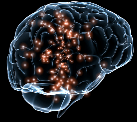
Neurons in nervous system communicate through synapses (a structure that allows neuron to transmit electrical or chemical signal to another neuron). This type of communication is called synaptic transmission or Neurotransmission that is fundamental to how the brain functions. The properties of synaptic connectivity and synaptic transmission between neurons are less known as their study is usually carried by using artificial conditions. It is very difficult to perform study of these transmissions in live animals in real time.
Now the study is possible as the researchers from Swiss Federal Institute of Technology in Lausanne (EPFL) performed a research in vivo (the effects of various biological entities are tested on living organisms mostly animals including humans). They measured synaptic transmission by using a technique that combined single-cell optogenetics with whole-cell recordings to investigate glutamatergic synaptic transmission in vivo. In other words, this study is the first of its kind where synaptic transmission was measured in a vivo by using a method that combines genetics and the physics of light to analyze specific neurons in real time. The light used also controled the activity of particular neurons.
Optogenetics is a technique in which a light-sensitive protein is inserted into living neurons. These modified neurons act as an electrical channel and produce light-sensitive protein that sits on the membrane. The channel opens up and allows the electrical ion to flow into the cell when the light falls on the neuron. This leads to optogenetic stimulus and if this stimulus is strong it will generate electrical signal in the neuron.
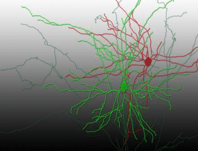
Aurelie Pala and Carl Petersen used optogenetics to stimulate neurons of anaesthetised mice. Pala focused on the neurons that were located barrel cortex of the mouse's brain. After showing blue light on neurons that consists of light-sensitive protein, the neurons got activated and fired signals. The advanced imaging technique known as two-photon microscopy was used. This technique helped them to study the brain of the live mouse and analyze the neurons.
You can have detailed look at the research that was published in the journal Neuron
Source: #-Link-Snipped-# via #-Link-Snipped-# | Image Credit: <a href="https://www.darpa.mil/uploadedImages/Content/NewsEvents/Releases/2014/SUBNET_Final_1.jpg" target="_blank" rel="nofollow noopener noreferrer">Defense Advanced Research Projects Agency - Content not found</a>
Now the study is possible as the researchers from Swiss Federal Institute of Technology in Lausanne (EPFL) performed a research in vivo (the effects of various biological entities are tested on living organisms mostly animals including humans). They measured synaptic transmission by using a technique that combined single-cell optogenetics with whole-cell recordings to investigate glutamatergic synaptic transmission in vivo. In other words, this study is the first of its kind where synaptic transmission was measured in a vivo by using a method that combines genetics and the physics of light to analyze specific neurons in real time. The light used also controled the activity of particular neurons.
Optogenetics is a technique in which a light-sensitive protein is inserted into living neurons. These modified neurons act as an electrical channel and produce light-sensitive protein that sits on the membrane. The channel opens up and allows the electrical ion to flow into the cell when the light falls on the neuron. This leads to optogenetic stimulus and if this stimulus is strong it will generate electrical signal in the neuron.
Aurelie Pala and Carl Petersen used optogenetics to stimulate neurons of anaesthetised mice. Pala focused on the neurons that were located barrel cortex of the mouse's brain. After showing blue light on neurons that consists of light-sensitive protein, the neurons got activated and fired signals. The advanced imaging technique known as two-photon microscopy was used. This technique helped them to study the brain of the live mouse and analyze the neurons.
You can have detailed look at the research that was published in the journal Neuron
Source: #-Link-Snipped-# via #-Link-Snipped-# | Image Credit: <a href="https://www.darpa.mil/uploadedImages/Content/NewsEvents/Releases/2014/SUBNET_Final_1.jpg" target="_blank" rel="nofollow noopener noreferrer">Defense Advanced Research Projects Agency - Content not found</a>